To enable
us to understand the possible mechanisms leading to "chronic prostatitis"
we have to know some basic facts about the anatomy and physiology of the
prostate.
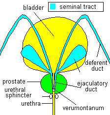
The prostate is a gland situated underneath the bladder (the bladder
neck) and is perforated by the first portion of the urethra.
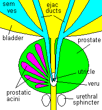
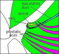
The 2 ejaculatory ducts enter the upper part of the prostate
from behind, travel through the gland and open into the urethra on a small
protuberance (3-4 mm) of the urethral mucosa called the verumontanum
("veru").
The veru is very critical because of the convergence of several other
structures:
- Between the 2 openings of the ejaculatory ducts, we find the opening
of the utricle, a remnant of our early life as embryo
(a small duct representing the rests of the tissue which in the female
develops into the uterus).
  The
appearance of the utricle can vary widely from a tiny dimple in the
veru to a long narrow duct extending deep into the prostatic tissue
parallel to the ejaculatory ducts in the midline. In some individuals,
this duct obliterates forming a cyst (utricular cyst) in the prostate,
not rarely the cause of symptoms identical to those of "chronic prostatitis". The
appearance of the utricle can vary widely from a tiny dimple in the
veru to a long narrow duct extending deep into the prostatic tissue
parallel to the ejaculatory ducts in the midline. In some individuals,
this duct obliterates forming a cyst (utricular cyst) in the prostate,
not rarely the cause of symptoms identical to those of "chronic prostatitis".
- The prostate is composed of 25-30 small glandular units (acini)
located in the periphery of the prostate, and each glandular unit is
connected to the outside world by a tiny duct which opens into the urethra
at each side in direct proximity to the veru. The prostatic acini produce
a fluid that, at orgasm, is expelled from the acini by contraction of
the prostatic smooth muscle tissue surrounding these acini. The composition
of the prostatic fluid is vital for the well-being of the spermatic
cells outside the body and severe alteration, like in certain forms
of chronic prostatitis, can degrade fertility.
Other important anatomical structures, mostly neglected in the literature,
are the seminal vesicles (SV). 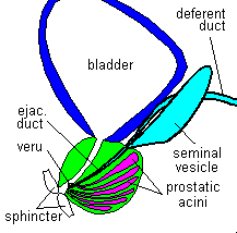 These
glands reside on the backside of the lower part of the bladder, their
body (about 5-8 cm long, .6-1 cm wide) lies alongside the deferent duct
(which carries the sperm cells from the testis to the urethra) and empties
into this duct before the deferent duct enters the prostate to become
the ejaculatory duct. The SVs are structurally hollow organs comparable
to the gallbladder, but with multiple small saccular compartments (looking
almost like a grape) interconnected with each-other. The wall of the
SVs is composed of an internal cellular lining (glandular cells) which
produces a fluid necessary for the extracorporeal survival of the sperm
cells. This fluid, together with the fluid from the prostatic acini,
constitutes a major part of the volume of the spermatic fluid; only
a small part comes from the testicles. The outside muscular shell of
the SVs contracts and expells the secretion at orgasm. These
glands reside on the backside of the lower part of the bladder, their
body (about 5-8 cm long, .6-1 cm wide) lies alongside the deferent duct
(which carries the sperm cells from the testis to the urethra) and empties
into this duct before the deferent duct enters the prostate to become
the ejaculatory duct. The SVs are structurally hollow organs comparable
to the gallbladder, but with multiple small saccular compartments (looking
almost like a grape) interconnected with each-other. The wall of the
SVs is composed of an internal cellular lining (glandular cells) which
produces a fluid necessary for the extracorporeal survival of the sperm
cells. This fluid, together with the fluid from the prostatic acini,
constitutes a major part of the volume of the spermatic fluid; only
a small part comes from the testicles. The outside muscular shell of
the SVs contracts and expells the secretion at orgasm.
In summary, in a minute spot of the prostatic urethra around the veru,
covering an area not larger then 1 square cm (about 1/6 square inch)
we find all the openings where the spermatic secretion has to pass through.
One can immagine that a slight change (focal inflammation with edema,
calcifications, microscars after inflammatory disease etc) can distort,
compress, obstruct (partially or completely, temporarily or definitively)
those tiny openings creating all the conditions necessary for disease
in one, few or many prostatic glandular subunits or the seminal tract.
Of course, if passage through one or several of these ducts is not completely
restored (e g due to inadequate treatment of an acute exacerbation of
prostatitis, permanent changes like calcified deposits of detritus or
scars) we'll have to expect chronification of the inflammatory process
(not always symptomatic) with acutisation from time to time. The close
relationship of the SVs and the prostate to the bladder neck and the
trigone (an area in the bladder floor adjacent to the bladder neck),
the most sensitive parts of the bladder with a dense concentration of
sensory nerve endings, explain the occurrence of urgent desire to void
frequently associated with irritative conditions in the prostate/SV.
In conclusion, an comprehensive appreciation of the anatomical structures
and their relationship is necessary for the understanding of the different
syndromes that run under the term of "chonic prostatitis", instead of
a view limited to the prostate only.
|
